Next Meeting: August 19 - Round Table. A panel of members talk of their experiences. Then the group will break-out into sessions by treatment type (Active Servaliance, Surgery, ADT, Radiation, Chemo) for networking.
June Meeting: GeorgeJohnson 20170717 - YouTube Hormone Therapy (also called ADT, Androgen Deprivation Therapy) An overview of ADT, its causes, perspective of urologists, controversies, and member experiences.
Our Last Meeting in July:
- Imaging and Genomics in Prostate Cancer Management
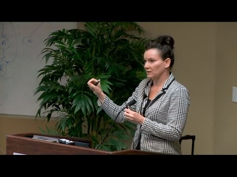
Bernadette started as a combat medic!
But she is a researcher, not a physician, and does not give medical
advice. She has a BS in radiologic
sciences, earned a postgraduate Certificate in Imaging Sciences from University
of Edinburgh and is working on a Ph.D. in tumor immunology imaging. Many awards and publications. A few years ago, she founded the
International Laser Network, a not-for-profit organization comprised of laser
users with a goal of keeping patients safe and educating users. She is a vocal activist for patient
care.
1. The history of biopsy
strategies: The first biopsies were done in the 1920's. Gradually, ultrasound guidance and "systematic"
biopsy grids were added, but they still miss significant tumors, even with
"saturation" biopsies using very many needles. A major advance occurred when MRI guidance
for biopsies was added (and officially recognized in about 2010), with
standardization now worked out.
Traditional screening for PCa (prostate cancer) is associated with
over-diagnosis and over-treatment of clinically insignificant PCa. Systematic TRUS (trans-rectal
ultrasound-guided) biopsy has a false negative rate of 30-35%, missing
clinically significant PCa. And systematic
TRUS biopsies under-estimate Gleason scores 30-40% because of missing the
most-significant tumors. Thus, clinical
staging based on TRUS biopsies underestimates pathological staging 15-25%. Furthermore, 26% of patients in active
surveillance harbor undetected clinically significant PCa (i.e., tumors that
probably should be treated without delay).
2. Technical aspects of MRI
imaging: Three main parameters are used to determine the likelihood of a
tumor being present in the imaged area:
T2 (see April 2017 IPCSG newsletter), DWI (diffusion of water is
restricted where cells are densely packed, which occurs commonly in tumors,
inflammation and infection), and DCE (dynamic contrast enhancement, looking at
the rate of MRI contrast (gadolinium-based contrast) entering and exiting the
tumor rapidly -- because of its higher-than-normal blood supply). According to the medical literature, together
they give better than 90% accuracy in detecting prostate tumors.
Functionally, a 1.5 Tesla magnet and a 3.0 Tesla magnet in the MRI unit
give the same results. Theoretically,
the 3.0 would be better, but air and movement in the pelvis wash out the
differences, so her group (and a local group, Imaging Healthcare Specialists)
prefers the 1.5 magnet. Data presented
at ASCO and AUA also support 1.5T for prostate cancer imaging as most scanners
in the U.S. are 1.5T. The main drawback
of 1.5T is that the sequences are slightly longer (seconds or minutes longer,
not hours).
Under current standardization, a multi-parametric MRI results in a
PI-RADS score, which indicates how abnormal the suspicious areas in the image
are. She feels the descriptors in the
table below could be more action oriented:
The classification labels should indicate whether a biopsy should be
performed, and at what level of urgency.
As a member of the ACR Pi-RADS subcommittee on imaging standards, she
has suggested the following language: 5=
biopsy immediately! 4= needs a biopsy. 3= probably doesn’t need a biopsy, but
wouldn’t hurt. 2= doesn’t need a biopsy. 1= don’t bother.

She described the procedure for MRI-guided biopsies – see the video. The procedure is fast: 20-30 minutes, and
very accurate. Some have promoted
“Fusion” biopsies, combining MRI images with real-time Ultrasound imaging, but
Bernadette explained that there is a plus-or-minus 3 mm inaccuracy (“skew in
the X-Y plane”) in published accounts of such biopsies, which she considers
unacceptable for biopsies of small tumors.
However, her office does accept results of such biopsies from some
experts in the technique, though her organization (Desert Medical Imaging) does
not do them.
Gleason scoring standards are changing, so it’s appropriate to know which
standard is used in your biopsy. Also,
consider getting a second opinion to reduce inter-observer variability in the
grading.
Biopsy samples should be sent for genomic testing – see discussion below.
3. Rationale for her early work (2008-2009)
on development and use of MRI-guided laser focal therapy of PCa: Of all options, she felt back then that Cryotherapy
and Laser therapy were the only two potential methods for focal therapy, but Cryo
gives much less control over the margins of the treatment – being more a
regional treatment than a precise, focal treatment.
In laser focal therapy, the laser fiber is inserted through the same
device as is used for biopsy, with a cooling catheter that only allows heating
at the tip of the laser fiber. An
interface allows creation of thermal maps from the MRI data, to precisely
monitor the treatment every 4 seconds.
After a low dose of heat from the laser, to confirm the tip placement, a
therapeutic treatment dose is given for up to 120 seconds, heating the tissue
to about 60-70° Celsius (140-160° F) to necrotize or kill the tumor. Safety cursors are placed on the image to
protect nearby structures, with the heating automatically cut off if the
temperature at those points gets to an unsafe level. The transition zone between treated and
unaffected areas is desirably very narrow, less than one millimeter – in
contrast to 5-10 mm in Cryotherapy, HIFU and Radiofrequency (RF) ablation.
4. Update on NCT #02243033 (Phase
II clinical trial of laser focal therapy):
Phase I safety and feasibility study results are to be published
soon. Little to no “morbidity” (side
effects). The rate of “positive margins”
was 26%, which is either due to some tumor left behind, or to recurrence at the
treatment site. Some tumor is often left
behind to avoid getting too close to other structures such as the urinary
sphincter, or when de-bulking a tumor that has extended into the seminal
vesicles or bladder wall in “salvage” patients (i.e., after some other
treatment).
On treatment naïve patients (i.e., no prior therapy), PSA scores
decreased by 35% at one year after laser focal therapy, with no statistically significant
change in IPSS (International Prostate Symptom Score for urologic function) or
SHIM (Sexual Health Inventory for Men) scores.
“Salvage” patients had 47% decrease in the mean PSA, and no
statistically significant change in the IPSS or SHIM scores (with 16 patients
so far).
Current conclusions about laser focal therapy are that it is feasible and
safe, with a recurrence rate of 25%, and about 5% going on to whole gland
therapy. Patients are still viable for
retreatment afterward, either for focal or whole gland therapy. There is a nice progression: Multiparametric MRI leads to MRI-guided
biopsy, which leads as needed to MRI-guided laser focal therapy in patients who
meet the study inclusion criteria.
The Phase II study (Phase I was converted in May 2016; also NCT #02243033)
is 7 years into a 20-year follow-up and these results were presented at the
American Association for Cancer Research, 2017.
5. PET (positron emission
tomography) imaging with Axumin imaging agent (recently approved for
commercial use; also known as FACBC):
The agent is suitable for immediate imaging, 3-5 minutes after injection,
and scanning takes 20-30 minutes.
Examples of imaging to pick up lymph node and other metastases were
shown. Other approved imaging agents are
F 18 FDG (low uptake by prostate cancer; bladder excretion obscures nearby
lymph nodes; used typically in patients with elevated PSA), F 18 NaF (used to
assess for bone metastasis), C 11 Choline (used after initial therapy, to
localize recurrence if rising PSA and inconclusive conventional imaging;
requires on-site cyclotron). Details on
studies and results are in the slides. Accuracy
with Axumin was about 80% in pelvic scans of salvage patients with PSA
>1.8. Adverse reactions were very low
and mostly mild. Half-life is 110
minutes, allowing use at sites that do not have a cyclotron.
6. Potential role of genomic
classifiers for risk stratification:
A biomarker is a measurement indicating normal or pathogenic biological
processes, or response to a therapy. For
prostate cancer, Desert Medical Imaging uses several non-invasive biomarker measurements,
including PSA and mpMRI (for PSA density, tumor volume, tumor staging, and
biopsy targeting) and uses MRI-guided biopsies for Gleason score and genomic
testing.
She inquired as to any present who had genomics done on their MRI-guided
biopsy? Only one.
There are many current and emerging genomic tests. Here are the most well-known:
ProstaVysion – 2 genes: MRG (overexpression of this gene is bad) and PTEN
(there are two versions of this helpful gene, but one or both may be
missing. Surprisingly, she finds that if
one PTEN is missing, it’s better, not worse, to have both missing according to
her current small data set, in contrast to ProstaVysion’s scoring system.)
ConfirmMDx – after negative biopsy, when cancer is still suspected, this
test is run using 8-18 cores.
Prolaris – 20-something genes are tested.
OncotypeDX – ditto.
Decipher – ditto. Until suggested
3 years ago by Bernadette, this test had only been done on prostatectomy
samples, but they now offer it for analysis of biopsy cores. Produces a 5-10 year metastatic risk profile. She now uses this on every patient where the
cores allow it, and with permission, submits their core samples for additional
genetic testing, up to 1.4 million genes in several ongoing research studies. Cores can be tested up to 7 years after the
biopsy.
Intratumoral and intertumoral heterogeneity of the prostate cancer
genomics has been shown, emphasizing the importance of targeted biopsies and
genomic testing for classification and prognostication of disease progression.
A final note: She recommends the NCCN.org patient prostate cancer treatment
guidelines, starting on page 45 of their pdf about prostate cancer, which is available
at https://www.nccn.org/patients/guidelines/cancers.aspx
Questions:
- PSMA agents? A member noted that they can be used at much lower PSA than Axumin. Bernadette is working to get 18F-DCFPyL as soon as possible – it is not commercially available yet, but is in a trial at Johns Hopkins. Gallium 68 is being used at UCSF -- 500 patients so far. Two group members have had good results there. Australia & Germany have been using Gallium 68 for many years. Other new agents were mentioned as being in active development.
- The immunological response (stimulation) after laser focal therapy? Not known yet, but she is studying tumor immunology imaging to work on it.
- Bottom line on laser focal therapy? “Oncological control without morbidity” -- but only studying its use on Gleason 7 or lower. The phase II trial will be completed 20 years after 1000 patients have entered the study.
- Cost? She has no grant money to help with costs. $25,000 entry fee to be treated. Desert Medical Imaging does not submit to insurance companies, but patients have sought reimbursement on their own. Men in financial hardship can apply for funding at www.thefocaltherpyfoundation.org, co-founded by Bernadette and her patient, Vinny Smith.
- Use of IBM’s Watson computer system? It will be a game-changer. She strongly favors the use of computers and automation in disease analysis and treatment planning, and is waiting for IBM to harvest her data.
- Cyberknife? Appropriate in some cases. She is in favor of: standardizing treatments, making them widely available, and using treatments that are the least damaging, the least traumatic, and that offer the highest hope for oncologic control. Seek advice from your physician.
Background/Related
- Synopsis of the PI-RADS v2 Guidelines for Multiparametric Prostate Magnetic Resonance Imaging and Recommendations for Use - European Urology
- The use of molecular imaging combined with genomic techniques to understand the heterogeneity in cancer metastasis
- In-bore magnetic resonance-guided transrectal biopsy for the detection of clinically significant prostate cancer
- Phase II Laser Focal Therapy of Prostate Cancer - Full Text View - ClinicalTrials.gov
- Axumin injection now available for PET imaging of recurrent prostate cancer - MedicalPhysicsWeb
- Beyond PSA: Prostate Cancer Biomarkers - wallen
- BCMJ_56_Vol7_PSA_prostate_cancer.pdf
- Genomic/genetic tests for risk for clinically significant prostate cancer | THE "NEW" PROSTATE CANCER INFOLINK
- A More Accurate Prostate Cancer Grading System
- Insights.New+Biomarkers+Tests+for+PC.Is16-3-2.pdf
No comments:
Post a Comment